

Human reproduction (AHL)
11.1 Production of gametes
11.1.1 Draw the structure of the testis as seen using a light microscope.
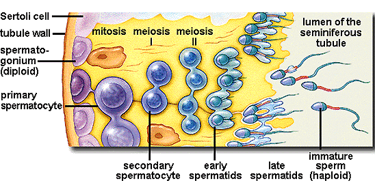
Light microscopes show the presence of seminiferous tubules with blood capillaries and interstitial cells. The sectioned tubules have an outer germ cell layer with basement membrane, developing spermatozoa and the Sertoli cells that provide nourishment.
11.1.2 Describe the process involved in spermatogenesis including mitosis, cell growth, the two divisions of meiosis and cell differentiation (cross reference 7.2, 7.3 and 10.1).
Mitosis produces the tubule germ cell layer, spermatocytes develop by cell growth which then undergo meiosis producing haploid spermatids that differentiate into spermatozoa.
11.1.3 Outline the origin and the role of the hormones FSH, testosterone and LH in spermatogenesis.
Limited to the hormones above. The name ICSH will not be used.
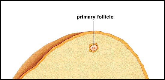
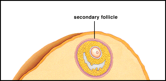
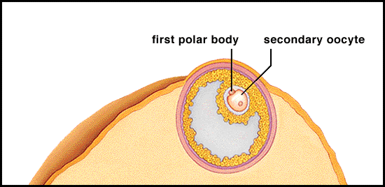
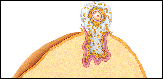
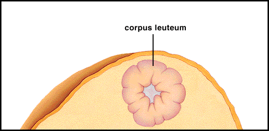
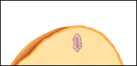
11.1.4 Draw the structure of the ovary as seen using a light microscope.
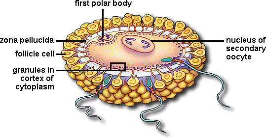
Developing oocytes can be seen. (Note: primary oocytes are made before birth). The stages of developing Graafian follicles are visible, the primary oocytes surrounded by the zona pellucida. Meiotic division I gives two haploid secondary oocytes with unequal cytoplasm and then ovulation occurs. Meiotic division II occurs after sperm penetration.
11.1. 5 Explain the process involved in oogenesis including mitosis, cell growth, the two divisions of meiosis and the unequal division of cytoplasm and the degeneration of polar bodies (cross reference 7.2 and 10.1).
Name of the stages are not required.
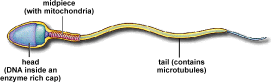
11.1.6 Draw the structure of a mature sperm and egg.
11.1.7 Outline the role of the epididymis, seminal vesicle and prostate gland in the production of semen.
11.1.8 Compare the process of spermatogenesis and oogenesis including number of gametes, timing of the formation and release of the gametes.
11.2 Fertilisation and pregnancy (2h)
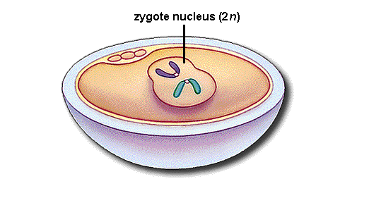
11.2.1 Describe the process of fertilisation including, the acrosome reaction, penetration of the egg membrane by a sperm, and the cortical reaction.
11.2.2 Describe the role of human chorionic gonadotrophin (HCG) in early pregnancy and pregnancy testing.
After implantation, the developing placenta makes HCG some of which is lost (with other hormones) in the urine. A monoclonal antibody to HCG is produced commercially. Although several commercial tests exist, many consist of dye molecules attached to monoclonal antibody in a plastic holder. When this is dipped into urine which has HCG the complex of hormone/monoclonal antibody/dye moves to appear a coloured line.
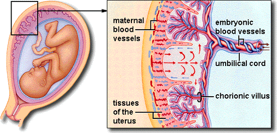
11.2.3 Describe the structure and functions of the placenta including, its hormonal role (oestrogen and progesterone) in the maintenance of pregnancy.
Details of the embryological development of humans, formation and evolutionary origins of the extra-embryonic membranes, hormonal control of lactation are not required. Prolactin in connection with 5.6.11 might also be discussed here.